Guide to Playing Mozak
Getting to Know the Project
- Introduction
- Getting to Know the Project
- Getting to Know a Neuron
- Getting to Know an Image Stack
- Noise vs. Signal
- Types of Neurons — Spiny/Aspiny
- Tracing Faint Branches
- Creating a Fully Connected Structure
- Creating Axon and Dendrite Traces
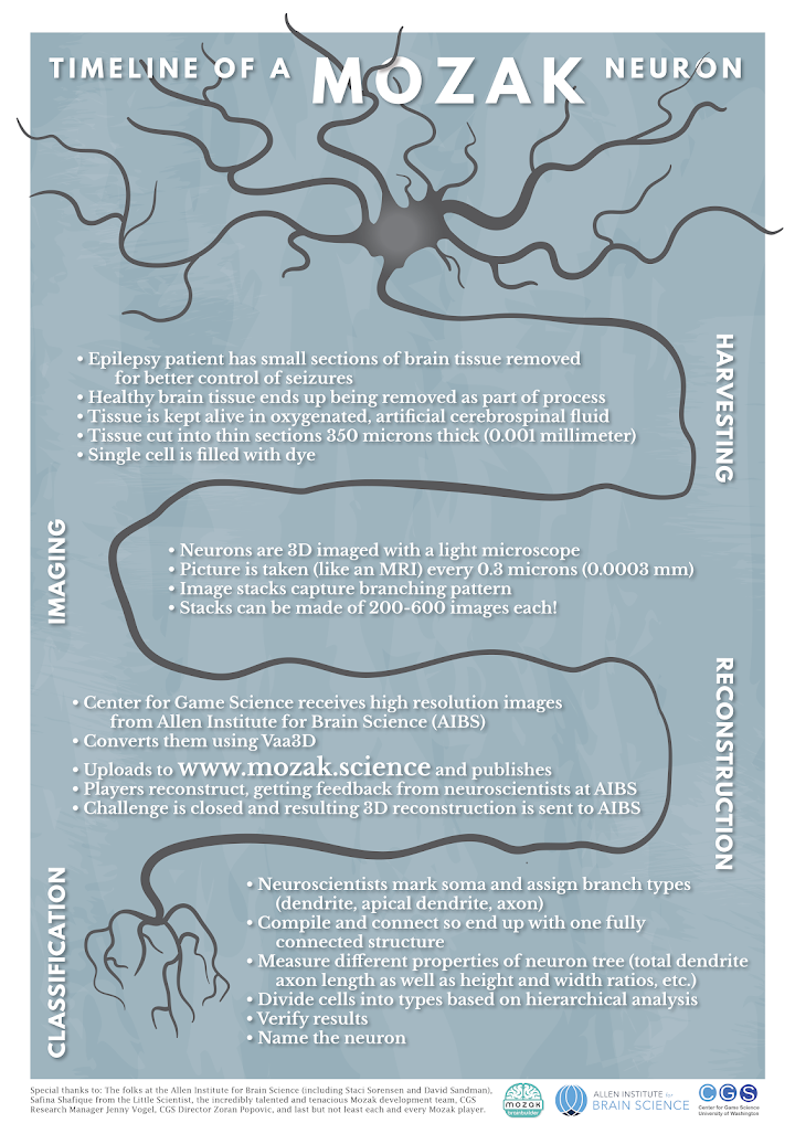
Each neuron is contained in a thin slice of brain tissue. A researcher at the Allen Institute for Brain Science places a small glass pipette in the tissue, and uses a technique called Patch-Clamp to record the electrophysiological profile of the neuron. The electrophysiological profile basically tells us how the neuron communicates to other neurons by measuring the strength and timing of its action potentials (among other things).
Before the pipette is removed from the tissue, it is used to fill the target neuron with a dye called biocytin that makes the neuron visible. The slice of tissue is then imaged at 63X magnification with a special microscope. The microscope takes hundreds of pictures of the tissue at different depths to create a 3D image stack (similar to how an MRI would work). Image stacks are posted on Mozak so players like you can reconstruct each of the labeled neurons. Your reconstructions capture the “morphology” of a neuron, which is just a fancy way of saying its shape. Our overall goal is to use the morphology and electrophysiological profile of each neuron to discover how many different types of neurons there are in the mouse visual cortex!
Check out the Cell Types Database being generated at the Allen Institute for Brain Science. This is the project your work will be assisting!