Guide to Playing Mozak
Getting to Know an Image Stack
- Introduction
- Getting to Know the Project
- Getting to Know a Neuron
- Getting to Know an Image Stack
- Noise vs. Signal
- Types of Neurons — Spiny/Aspiny
- Tracing Faint Branches
- Creating a Fully Connected Structure
- Creating Axon and Dendrite Traces
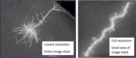
When you load a neuron on Mozak, the first thing you will see is the whole image stack. From this initial view, the cell will appear pixelated. This is because the full resolution image stacks are massive; they can be over 100GB!! We have to down-sample them to show you the whole image stack, otherwise the loading times would be unbearable. If you use the zoom function in the upper-left corner of the window, you will notice that the resolution of the cell improves as the area shown gets smaller. Here is an example:
It is most efficient to find a compromise between resolution and visible area. You will want to have enough resolution to be able to trace accurately, but enough visible area that you will not have to pan every few seconds.
The 3D image stack is made up of hundreds of 2D images stacked on top of one another. To see a single plane, you will need to use the connect the dots tool.
Tracing from the whole stack view using the virtual finger tool is appropriate for capturing thick, well-labeled neurites (usually dendrites), while tracing from the single plane view using the connect-the-dots tool is often used for faintly-labeled neurites (usually axon). When you are in the single plane view, use your mouse wheel to scroll between sections!
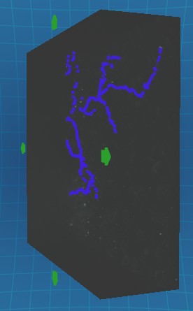
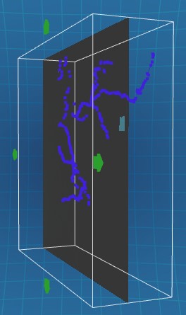
The last thing to know about 3D image stacks is that they have different zones, and in each zone the neurites will look a little different. Below is a side view of an image stack separated by zone with the left side being closest to the viewer.
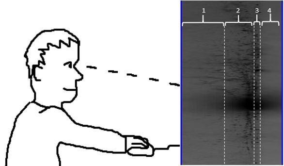
- Zone one contains the planes that are closest to the viewer. These are also the deepest planes in the brain slice. Because they are deep, the signal here is usually much harder to see. All neurites (dendrites and axon) will appear faint and more diffuse. Boost the brightness slider to find all the signal here!
- Zone two contains planes that are shallow in the slice, where the signal is easily seen and captured. The soma is almost always in zone two.
- Zone three is very thin and contains the planes at the surface of the slice. The thing to know about this zone is that there is usually a bunch of false signal on the surface of the slice. Try to avoid tracing here unless you see a continuous, easily identifiable neurite.
- Zone four contains the planes furthest from the viewer. These planes are images taken above the surface of the slice, which means they will not contain any signal or tissue at all. Essentially, the microscope focused on the air above the slice. Note that this zone may be much thinner than in the example above.